Mild traumatic brain injury (mTBI) is among the most commonly under-documented neurological conditions in clinical and legal practice not because the injury is rare, but because the standard diagnostic tools used to evaluate it were never designed to detect it.
Understanding that gap is essential for any clinician managing post-concussive patients, and for any attorney handling personal injury cases involving head trauma.
The Standard Workup and Its Limits
In the acute setting, the standard first-line imaging for suspected TBI is a non-contrast CT scan. It is fast, widely available, and excellent at identifying what it was designed to find: intracranial hemorrhage, skull fractures, cerebral edema, and major structural lesions.
But mild traumatic brain injury rarely produces those findings. The injury mechanism in mTBI is fundamentally different, it operates at a microscopic level, within the white matter architecture of the brain, well below the resolution threshold of CT and often of conventional MRI as well.
The numbers are unambiguous: studies consistently show that 85 to 99 percent of adults presenting with mild TBI will have a normal CT scan. A normal result does not rule out injury. It rules out the specific findings CT was built to detect.
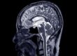
Standard MRI sequences – T1, T2, FLAIR – improve upon CT in sensitivity for certain findings, but still fall significantly short when it comes to the diffuse axonal injury (DAI) that characterizes most mTBI. Axonal disruption at the microstructural level produces no visible lesion on conventional sequences. The scan appears normal. The patient is not.
The Underlying Pathophysiology
In mTBI, injury most commonly results from rotational acceleration-deceleration forces – the rapid shifting and twisting of brain tissue within the skull that occurs during vehicular impact, whiplash, falls, or blunt head trauma.
These forces place mechanical stress on axons: the long, slender nerve fibers that form the white matter tracts connecting cortical and subcortical regions. When axons are stretched or sheared beyond their tolerance, the result is diffuse axonal injury – disrupted signal transmission across distributed neural networks.
The clinical presentation reflects this distributed disruption: slowed processing speed, working memory deficits, executive dysfunction, emotional dysregulation, fatigue, and post-concussive headache. Symptoms that are real, measurable on neuropsychological testing, and functionally significant, yet invisible on standard imaging.
Where DTI Fits
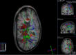
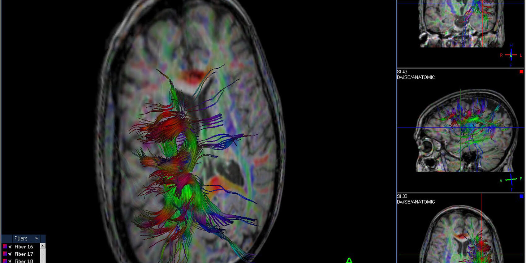
Diffusion Tensor Imaging (DTI) is an advanced MRI technique that measures the directional movement of water molecules through brain tissue. In intact white matter, water diffuses preferentially along the axis of healthy axons – a property called anisotropy. When axonal integrity is compromised, diffusion becomes less directional, more disorganized.
The primary DTI metric, Fractional Anisotropy (FA), quantifies this directionality. Reduced FA values in specific white matter tracts – particularly the corpus callosum, superior longitudinal fasciculus, frontal projections, and cingulum have been consistently associated with mTBI in the research literature, often in patients with normal conventional imaging.
Additional metrics including Mean Diffusivity (MD), Axial Diffusivity (AD), and Radial Diffusivity (RD) provide complementary information about the nature and extent of white matter disruption.
Critically, DTI does not diagnose TBI in isolation. Findings must be interpreted in the context of clinical history, neurological examination, symptom presentation, and neuropsychological data. What DTI provides is objective, quantifiable imaging data that can corroborate or contextualize a clinical picture that standard imaging cannot address.
Clinical and Medicolegal Relevance
For the treating clinician, DTI findings can inform treatment planning, support referral decisions, and provide objective documentation of white matter pathology in patients with persistent post-concussive syndrome.
For the personal injury attorney, DTI offers something that has historically been difficult to obtain in mild TBI cases: objective imaging evidence. In cases where a client presents with significant functional impairment and a stack of normal CT and MRI reports, advanced neuroimaging can provide a documented, radiologically-grounded basis for the injury claim.
Courts and medical experts have increasingly recognized DTI as a clinically meaningful tool. Its value lies not in serving as a standalone diagnosis, but in providing objective data that bridges the gap between a patient’s reported symptoms and the documented neurological findings that support them.
Advanced DTI Imaging at Kansas City Advanced Imaging
Kansas City Advanced Imaging (KCAI) provides advanced 3T MRI neuroimaging including DTI protocols designed to support the comprehensive evaluation of traumatic brain injury and post-concussive pathology.
KCAI’s platform is built around high-resolution 3T acquisition, advanced white matter imaging capability, and rapid outpatient access serving both clinical referral and medicolegal documentation needs in the Kansas City region.
For patients presenting with persistent post-concussive symptoms and unremarkable conventional imaging, DTI evaluation through KCAI may provide the objective neuroimaging data that the clinical picture warrants.
Kansas City Advance Imaging
Get In. Find Out.™
At KCAI, we believe meaningful change starts with the right support, guidance, and environment. Whether you’re exploring new paths for personal growth, seeking deeper understanding, or looking for solutions tailored to your unique journey, our team is here to help you move forward with confidence.
Every step you take matters, and we’re committed to helping you make the most of it. Reach out today to learn how we can support your goals and help you create lasting, positive change.
Unlike many imaging clinics, we offer same-day and next-day MRIs–schedule yours today.