If you’ve been told you might benefit from a DTI scan or if you’ve heard the term and wondered what it actually means, this is for you.
DTI stands for Diffusion Tensor Imaging. It’s a specialized form of MRI. And while the name sounds technical, the idea behind it is surprisingly intuitive once you understand what it’s measuring and why it matters after a head injury.
Start With the Brain’s Wiring
Most people know that the brain has gray matter, the tissue responsible for thinking, memory, emotion, and processing. But the brain also has an enormous amount of white matter, and that’s where DTI becomes important.
White matter is the brain’s communication network. It’s made up of millions of axons – long, slender nerve fibers that carry electrical signals from one part of the brain to another. Think of them like fiber optic cables running through a building: individually tiny, but collectively responsible for every signal that travels through the system.
These axons are bundled together into pathways called white matter tracts. Different tracts connect different regions – some link the two hemispheres of the brain, some connect the frontal lobe to memory centers, others support attention, emotional regulation, and coordination.
When those pathways are working well, information moves quickly and efficiently. When they’re disrupted (even at a microscopic level) communication slows down, gets scrambled, or breaks down in ways that can produce very real neurological symptoms.
What Happens to White Matter in a Brain Injury
In a mild traumatic brain injury, the most common mechanism of damage isn’t a direct blow to the skull – it’s the rotational force that happens when the head snaps suddenly. A car accident. A fall. A sports collision. A whiplash event.
That rapid rotation causes the brain to shift and twist inside the skull. The gray matter – the denser, heavier tissue – moves at a different rate than the white matter. That difference in motion stretches and strains the axons, sometimes to the point of microscopic tearing or disruption.
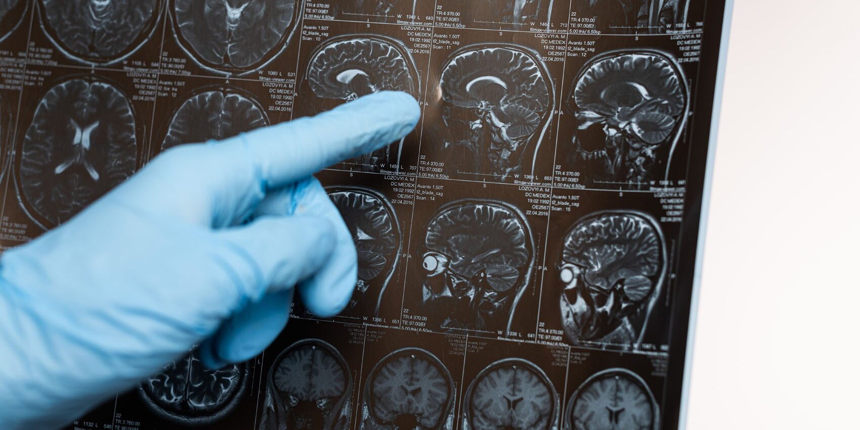
This is called diffuse axonal injury. It’s “diffuse” because it tends to affect axons across multiple regions rather than in one concentrated spot. And it’s invisible on a conventional CT or standard MRI because those scans aren’t designed to see changes at the microscopic level of individual axons.
This is the core reason so many mild TBI patients have normal imaging and real symptoms at the same time. The injury happened. The scans just couldn’t find it.
How DTI Works
Diffusion Tensor Imaging takes a different approach. Instead of looking at brain structure the way conventional MRI does, DTI measures the movement of water molecules through brain tissue.
Here’s the key insight: in healthy, intact axons, water doesn’t move randomly. It moves preferentially along the length of the fiber in an organized, directional way. Scientists call this anisotropy, meaning “direction-dependent.”
When axons are damaged or disrupted, that orderly movement breaks down. Water starts diffusing in less organized patterns – more randomly, less directionally.
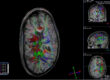
DTI detects and maps those diffusion patterns across the entire brain. The primary measurement it produces is called Fractional Anisotropy, or FA. Higher FA values indicate more organized, intact white matter. Lower FA values in specific regions may suggest disruption – potential axonal injury in those pathways.
The result is a detailed map of white matter integrity across the brain, capable of revealing abnormalities that no conventional scan could detect.
What DTI Can, and Can’t Tell You
DTI is a powerful tool, but it’s important to understand what it is and isn’t.
It is not a standalone diagnosis. A DTI scan doesn’t automatically confirm a traumatic brain injury or determine its cause. Findings must always be interpreted alongside clinical history, symptom assessment, neurological examination, and physician judgment.
What it can do is provide objective imaging data about white matter integrity – data that may corroborate symptoms that have gone unexplained, and that may be part of a broader neurological evaluation that helps complete the picture.
For many patients, DTI is the first imaging test that reflects what they’ve been experiencing. That matters – both clinically, and personally.
Why Scan Quality Matters
Not all DTI is created equal. The quality of a DTI scan depends heavily on the strength of the MRI scanner, the imaging protocols used, and the expertise of those interpreting the results.
Advanced 3T MRI systems – like those at Kansas City Advanced Imaging (KCAI) – provide significantly better signal quality and white matter resolution than lower-strength systems. DTI abnormalities in mild TBI tend to be subtle. Higher-quality imaging gives those subtle findings the best chance of being detected and accurately interpreted.
If you’ve experienced a head injury and are living with persistent symptoms, advanced neuroimaging may be an important part of understanding what’s happening – and what comes next.
Kansas City Advance Imaging
Get In. Find Out.™
At KCAI, we believe meaningful change starts with the right support, guidance, and environment. Whether you’re exploring new paths for personal growth, seeking deeper understanding, or looking for solutions tailored to your unique journey, our team is here to help you move forward with confidence.
Every step you take matters, and we’re committed to helping you make the most of it. Reach out today to learn how we can support your goals and help you create lasting, positive change.
Unlike many imaging clinics, we offer same-day and next-day MRIs–schedule yours today.