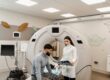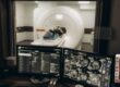
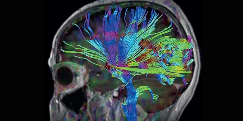
The human brain contains billions of neurons communicating through complex electrical and chemical signals every second. Understanding how different regions of the brain function has long been one of medicine’s greatest challenges. Functional Magnetic Resonance Imaging, commonly known as fMRI, has transformed neuroscience by allowing researchers and physicians to observe brain activity as it happens.
Unlike traditional MRI scans that focus primarily on anatomy and structure, functional MRI provides insight into how the brain is working in real time. This advanced imaging technology has become an invaluable tool for neurological research, surgical planning, and understanding a wide range of brain disorders.
What Is Functional MRI?
Functional MRI is a specialized MRI technique that measures changes in blood flow throughout the brain. When a particular area of the brain becomes active, it requires more oxygen and nutrients to support the increased workload of its neurons.
The body responds by increasing blood flow to that region. Functional MRI detects these subtle changes in oxygen-rich and oxygen-poor blood, creating detailed maps that show which parts of the brain are active during specific tasks.
This process is known as Blood Oxygen Level Dependent (BOLD) imaging, which serves as the foundation of most modern fMRI studies.
How Brain Activity Appears on an fMRI Scan
During an fMRI examination, patients may be asked to perform simple tasks while inside the MRI scanner. These activities could include:
- Reading words
- Listening to sounds
- Moving a hand or foot
- Viewing images
- Solving simple problems
- Answering questions
As the patient performs these tasks, the scanner continuously captures images of the brain. Advanced computer software analyzes changes in blood oxygen levels and identifies regions experiencing increased activity.
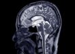
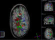
The resulting images display areas of activation as color-coded overlays placed on detailed anatomical MRI images. These maps help physicians visualize which brain regions are involved in specific functions such as language, memory, movement, or sensory processing.
Why Real-Time Brain Mapping Matters
One of the greatest advantages of functional MRI is its ability to identify critical brain functions before surgery or treatment.
For patients undergoing brain surgery, preserving important functional areas is essential. Functional MRI can help locate regions responsible for:
- Speech and language
- Motor control
- Vision
- Memory
- Sensory processing
Surgeons can use this information to plan procedures that minimize risks to critical brain functions while effectively treating tumors, epilepsy, or other neurological conditions.
This personalized mapping provides valuable information that may not be visible on conventional imaging alone.
Applications Beyond Surgical Planning
Functional MRI has become a powerful research tool for studying how the brain operates in health and disease. Researchers use fMRI to investigate conditions such as:
- Alzheimer’s disease
- Parkinson’s disease
- Stroke
- Epilepsy
- Traumatic brain injury
- Depression
- Anxiety disorders
By observing patterns of brain activity, scientists can gain a deeper understanding of how these conditions affect neural networks and brain function.
Functional MRI has also contributed significantly to research involving learning, memory, decision-making, emotional regulation, and cognitive performance.
The Role of Advanced MRI Technology
Functional MRI requires sophisticated imaging equipment capable of producing highly detailed images while detecting extremely subtle changes in blood oxygen levels. Advanced MRI systems provide the image quality, speed, and precision necessary for accurate brain mapping.
Higher-resolution imaging improves the ability to identify activation patterns and distinguish between neighboring brain structures. This enhanced accuracy supports both clinical decision-making and scientific research.
As imaging technology continues to advance, functional MRI is helping physicians and researchers unlock new insights into the brain’s remarkable complexity. By mapping brain activity in real time, fMRI offers a powerful window into how we think, move, communicate, and experience the world around us.
Kansas City Advance Imaging
Get In. Find Out.™
At KCAI, we believe meaningful change starts with the right support, guidance, and environment. Whether you’re exploring new paths for personal growth, seeking deeper understanding, or looking for solutions tailored to your unique journey, our team is here to help you move forward with confidence.
Every step you take matters, and we’re committed to helping you make the most of it. Reach out today to learn how we can support your goals and help you create lasting, positive change.
Unlike many imaging clinics, we offer same-day and next-day MRIs–schedule yours today.