Most concussions resolve. The headache fades, the fogginess lifts, and within a few weeks life returns to normal.
But for a significant number of people, that doesn’t happen.
Weeks pass. Then months. The symptoms linger – or come and go unpredictably. You’ve seen your doctor. You’ve had the scans. You’ve been told to rest, to be patient, that it will get better.
And still, something isn’t right.
If that’s where you are, this post is for you.
When Concussion Symptoms Don’t Follow the Script
Post-concussive syndrome is a recognized medical condition, persistent neurological symptoms following a mild traumatic brain injury that last beyond the typical recovery window. Estimates vary, but somewhere between 15 and 30 percent of concussion patients experience symptoms lasting longer than a month, and for some, symptoms persist for a year or more.
Those symptoms can include:
Persistent headaches or pressure in the head. Cognitive fog – difficulty thinking clearly, finding words, or following complex conversations. Memory problems, both short-term and with retention. Fatigue that isn’t explained by sleep. Irritability or emotional changes that don’t feel like “you.” Sensitivity to light or sound. Dizziness or balance issues. Anxiety or depression. Difficulty concentrating at work or school.
These aren’t character flaws or exaggerations. They’re neurological symptoms. And they deserve neurological evaluation – including the most advanced imaging available.
Why Your Previous Imaging May Not Be Enough
If you’ve already had a CT scan or a standard MRI and been told everything looks normal, that result is real – but it’s not the whole story.
Standard imaging is designed to detect structural damage: bleeding, swelling, fractures, large lesions. It does that well. What it cannot do is visualize the microscopic white matter injury that underlies many cases of persistent post-concussive syndrome.
The type of damage that causes lingering symptoms, diffuse axonal injury occurs at the level of individual nerve fibers, far below the resolution of conventional CT or MRI. A normal scan doesn’t mean the brain is uninjured. It means the injury is smaller than what those tools were built to find.
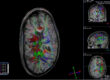
Advanced neuroimaging techniques, particularly Diffusion Tensor Imaging (DTI), were specifically developed to address this gap.
Who Should Consider Advanced Neuroimaging
DTI evaluation may be worth discussing with your physician if you:
Are still experiencing symptoms more than four to six weeks after a concussion or head injury. Had a head injury in a vehicle accident, fall, sports collision, or any event involving sudden impact or whiplash. Have had normal CT or MRI results but continue to have cognitive, neurological, or emotional symptoms. Are involved in a personal injury or workers’ compensation case where objective documentation of injury is important. Have been told your symptoms are “stress” or “anxiety” but believe they began with or worsened after a head injury.
DTI is not appropriate for every patient in every circumstance. Your physician is the right person to determine whether advanced imaging is indicated in your specific case. But knowing that it exists – and knowing what it can detect – means you can have an informed conversation about your options.
What the Process Looks Like
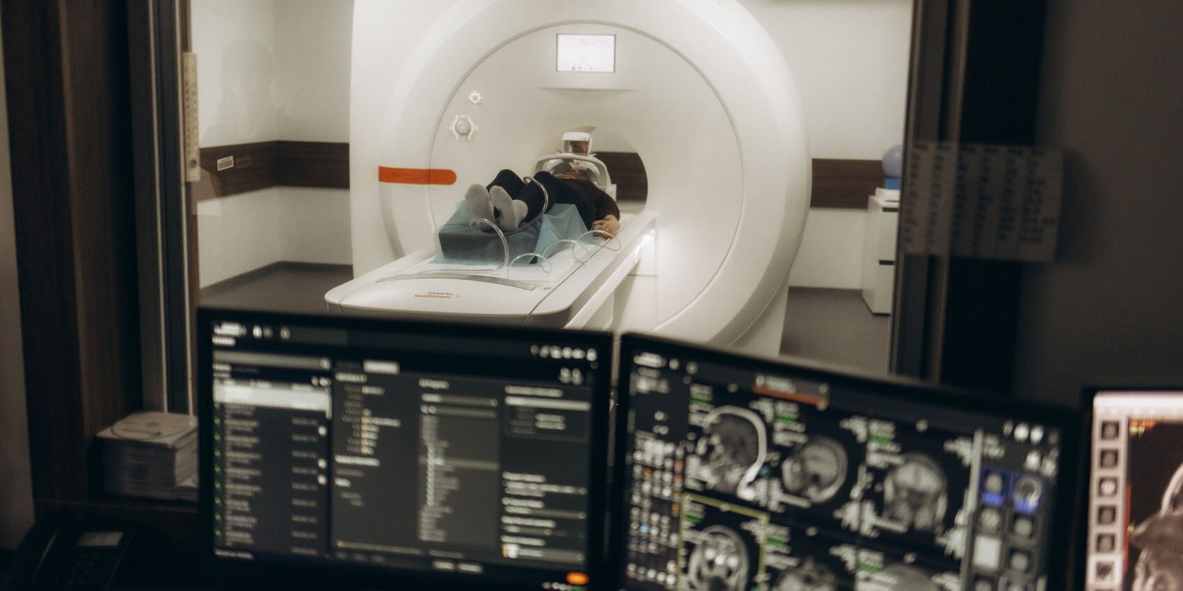
A DTI scan is performed on an MRI machine, the same kind of enclosed scanner you may have experienced before. For DTI specifically, a 3T (3 Tesla) system is strongly preferred, as the higher magnetic field strength produces significantly better image quality and white matter resolution.
The scan itself typically takes between 45 minutes and an hour. There’s no radiation involved. You’ll need to stay still during the acquisition, as motion can affect image quality. Most outpatient imaging centers can accommodate the scan without a lengthy wait.
After imaging, the DTI data is processed and interpreted by a radiologist with expertise in advanced neuroimaging. Results are typically shared with your referring physician, who integrates the findings with your clinical history and other test results.
DTI findings don’t produce a simple “positive” or “negative” result, they provide quantitative data about white matter integrity that your physician uses as part of a broader evaluation. The goal is a more complete picture, not a single definitive answer.
Taking the Next Step
Kansas City Advanced Imaging (KCAI) offers advanced 3T MRI neuroimaging including DTI protocols designed for the evaluation of traumatic brain injury and persistent post-concussive symptoms.
KCAI’s outpatient model is built around rapid scheduling access, high-resolution imaging technology, and a patient-centered experience. Whether you were referred by a physician, a specialist, or are exploring options on your own, KCAI can be a resource in your path toward answers.
You don’t have to keep explaining symptoms that no one has been able to document. Advanced neuroimaging exists precisely for situations like yours.
Kansas City Advance Imaging – Get In. Find Out™.
At KCAI, we believe meaningful change starts with the right support, guidance, and environment. Whether you’re exploring new paths for personal growth, seeking deeper understanding, or looking for solutions tailored to your unique journey, our team is here to help you move forward with confidence.
Every step you take matters, and we’re committed to helping you make the most of it. Reach out today to learn how we can support your goals and help you create lasting, positive change.
Ask your physician about referring to Kansas City Advanced Imaging for Same Day or Next Day MRI scheduling. Schedule yours today.